 One app for all your study abroad needs


The 7th chapter of the Biology class 11 syllabus, Structural Organisation in Animals, help us in understanding the formation of the structure in all complex animals. This topic familiarises students with the different types of functions that the animals are capable of performing. Thus, we have simplified the topic with examples that will make your understanding of the topic even better. So let us begin the tour of this topic.

This Blog Includes:
Animal Tissues
Tissues are nothing but an ensemble of homogenous cells carrying out certain similar functions. For classification, tissues of complex multicellular animals fall under four categories, namely, Epithelial, Connective, Muscular, and Neural.
Epithelial Tissue
Epithelia are compactly packed cells that look like an intracellular matrix and provide a lining to different body parts. Epithelial tissues are classified into two types such as simple epithelium and compound epithelium. Let us take a detailed look at these
Simple Epithelium
Simple epithelium provides lining to cavities, ducts, and tubes in the body. The simple epithelium is made up of a single layer of cells. Structurally, the simple epithelium is further divided into the following:
- Squamous Epithelium: Single thin layer of flattened cells lined with irregular boundaries makes for the Squamous epithelium. Their main function is to form a diffusion boundary. They are present in blood vessel walls and air sacs of the lungs
- Cuboidal Epithelium: Single layer of cube-like looking cells make up the cuboidal epithelium. They are present in the ducts of glands and a tabular section of nephrons of the kidney. Their main function is absorption and secretion
- Columnar Epithelium: Columnar epithelium have tall and slender cells with nuclei at its base. They can be located in the lining of the stomach and intestine and perform the function of secretion and absorption
Accorrding to structural organisation in Animals, when the columnar or cuboidal cells get specialised for selection, they form a Granular Epithelium which can be unicellular (isolated goblets in the alimentary canal) or multicellular (salivary glands).
| Exocrine Glands | Endocrine Glands |
| -They secrete substances such as mucus, saliva, earwax, oil, milk, digestive enzymes, and products of cells -Released through ducts or tubes. | -They secrete a compound called hormones. -Released directly as a fluid into the gland. |
Compound Epithelium
They are multi-layered and play an insignificant role in secretion and absorption. Their role is to protect the cells against chemical and mechanical stress.
Connective Tissues
They support and link other tissues in the body and are plenty in number. They are classified into three types:
- Loose Connective Tissues: Present beneath the skin with loosely arranged fibres (areolar tissue)
- Dense Connective Tissues: They consist of densely packed fibres. The collagen fibres are parallelly positioned between a cluster of fibres like tendons and ligaments
- Specialized Connective Tissues: Cartilage, bones, and blood, are types of specialized connective tissues
Muscle Tissues
The movement of the body requires the muscles to function by contracting (shortening) and relaxing (lengthening) the muscle fibres in response to stimulation. As mentioned in the chapter structural organisation in Animals, there are three types of muscles:
- Skeletal Muscle: The tissues are closely compressed to the skeletal bones in a parallel fashion. The biceps are made up of skeletal muscles
- Smooth Muscle: They are bundled together, taper at both ends, and are involuntary in nature. They can be found in the walls of organs such as blood vessels, stomach, and intestine
- Cardiac Muscle: It is present only in the heart; the cells contract in unison through fused plasma membranes

Neural Tissues
They control the responses of the body under different conditions. The neurons are protected by the neural system. The stimulation of neurons leads to electrical disturbance. When this disturbance is carried by the plasma membrane, they cause further disturbance to other cells.
In the coppming topic of our notes of structural organisation in animals is regarding the anatomy and morphology of earthworm, frog, and cockroach.
Click Here for Structural Organisation in Animals NCERT PDF
Earthworm
As per the chapter, here are some of the important pointers regarding the morphology and anatomy of earthworm-
- In India, we can find earthworms such as Pheretima and Lumbricus. They are reddish-brown and are found in moist soil. Its cylindrical shaped body is divided into over a hundred identical segments and is covered by a thin layer of cuticle
- The prominent regions of the body are segmented as prepatellar, clitellar, and postclitellar
- Its upper body segment is called peristomium and consists of the mouth area
- They don’t have lungs and breathe through their skin by dissolving oxygen from the moist surfaces they stay in
- Thin long tubes called nephridia perform the excretory processes that consist of three parts: nephrostome, the body, and terminal duct
- Ganglia is present in its nervous system and is divided into the central, peripheral, and sympathetic nervous system
- Their receptor cells help them to sense light and feel the vibrations of the ground under them. The chemoreceptors (taste receptors) absorb chemical information from the environment
- The earthworm is hermaphrodite and consists of both female and male organs. Sperm is exchanged during mating, which happens when two worms line up inverted to each other
Frogs
The details about the anatomy and morphology of frogs from the chapter structural organisation of animals are as follows-
- Frogs are Amphibia and are capable of surviving on land and in freshwater. In India, the Rana tigrina species of frog is commonly found
- Being cold-blooded animals, frogs tend to change their body temperatures according to the environmental temperature
- Frogs change their colour depending on the surface they are sitting on and camouflage to protect themselves from enemies
- Their skin feels slippery as it is covered in mucus. They stay in moist areas and absorb water through their skin to keep themselves hydrated
- The oxygen enters the frog’s body through its skin while it is underwater. On land, the frog uses its skin, lungs, and buccal cavity to breath
- The frog has a combined reproductive and excretory system referred to as the urogenital system. The frog uses a pair of kidneys, ureters, urinary bladder, and cloaca to eliminate waste from its system
- The frog has developed sensory organs for smell (nostrils), taste (tongue), hear (eardrums called as tympanum), sight (large protruding eyes), and touch (skin with lateral receptors)
- At a time, female frogs expel 2,500 to 3,000 eggs during mating. The fertilization process happens in water, and after 2 to 9 days, a tadpole is born

Cockroach
[optin-monster-shortcode id=”xf2mlnjiouddzrshykdb”]Mentioned below are the key pointer derived from the anatomy and morphology of Cockroach-
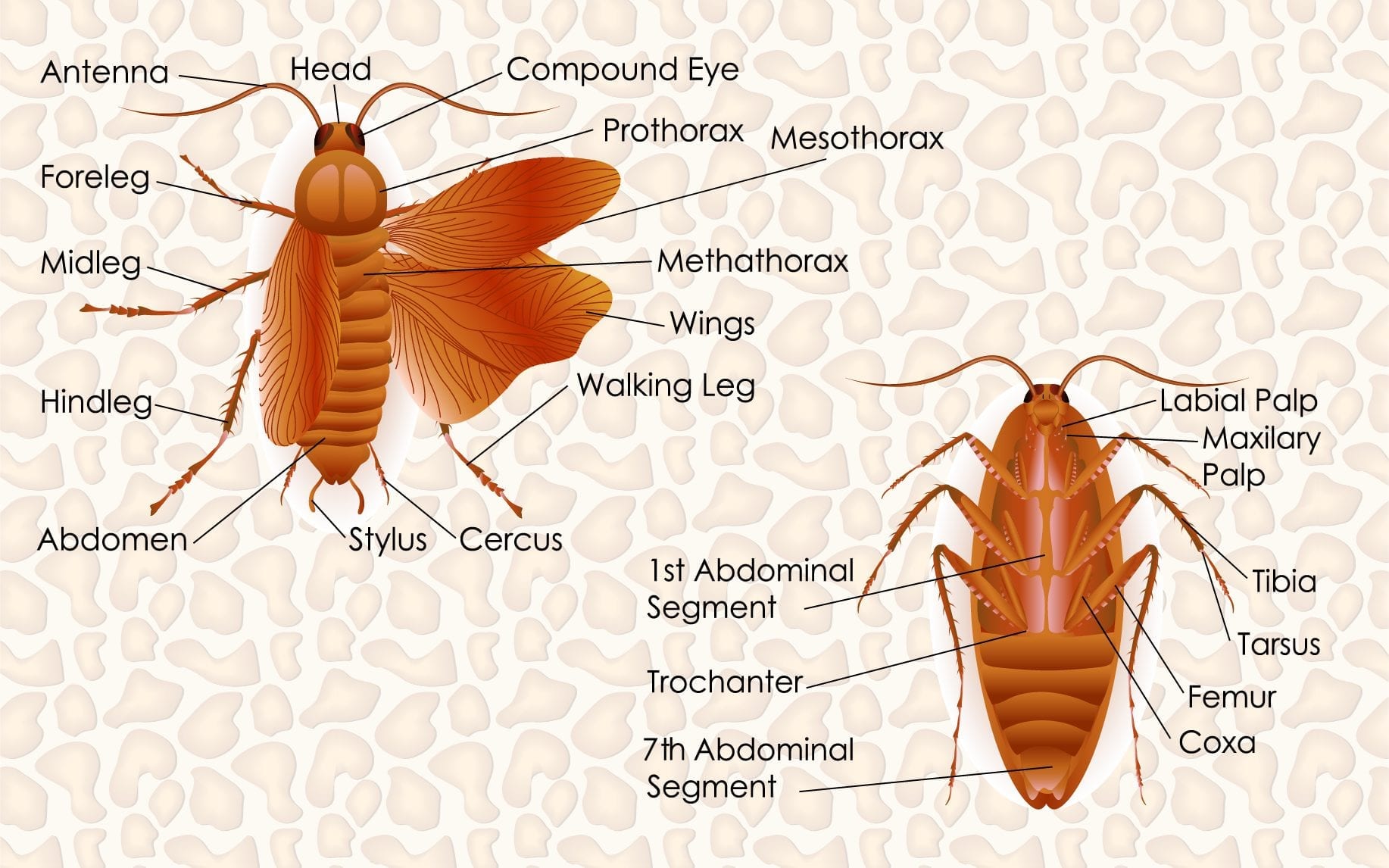
- Cockroaches belong to the Phylum Arthropoda family, and their bodies are divided into three segments: head, thorax, and abdomen. The body is engulfed by a hard exoskeleton made up of plates called sclerites
- Its triangular-shaped head (comprising six segments) has excellent mobility which is offered by its flexible neck. At either side of its head capsule are kidney-shaped compound eyes, out of which the antenna protrudes. The antennae act as sensory receptors and absorb information from the surroundings
- The respiratory organ of cockroaches is called tracheae which has ten small openings called spiracles on the lateral sides of the body
- Cockroaches excrete waste through the Malpighian tubules which convert the waste to uric acid, and hence, cockroaches are uricotelic insects
- Both male and female cockroaches have well developed reproductive organs. The female cockroach collects their eggs in a hard oval capsule called ootheca. Most females deposit their ready to hatch eggs near a crevice that has access to some food
- Most cockroaches are of no particular importance. These pests spread diseases by contaminating food with their excreta
Structural Organisation in Animals PPT
Structural Organisation in Animals Important Questions
- Explain the respiratory organ of cockroach
- Explain circulatory system of earthworm with the help of Diagram
- Briefly explain about Malpighian tubules
- Name all fuctions of Nephridia
- What are Neural tissues?
Structural Organisation in Animals Class 11 NCERT Solutions
(i) Give the common name of Periplanata americana
(ii) How many spermathecae are found in earthworm?
(iii) What is the position of ovaries in cockroach?
(iv) How many segments are present in the abdomen of cockroach?
(v) Where do you find Malpighian tubules?
(i) American Cockroach
(ii) 4 Pairs
(iii) Two Overies
(iv) 10 Segments
(v) Malpighian tubules are found at the junction of midgut and hindgut in the alimentary canals of insects

Credits: Pinterest
The cellular components include white blood cells, red blood cells and platelets.
(a) Areolar tissue; blood; neuron; tendon
(b) RBC; WBC; platelets; cartilage
(c) Exocrine; endocrine; salivary gland; ligament
(d) Maxilla; mandible; labrum; antennae
(e) Protonema; mesothorax; metathorax; coxa
(a) Neuron
(b) Cartilage
(c) Ligament
(d) Antennae
(e) Protonema
We hope that you found these notes of chapter structural organisation in animals interesting. Get in touch with our experts at Leverage Edu and they will guide you in getting directed towards your dream career path. Book and e-meeting now!

 45,000+ students realised their study abroad dream with us. Take the first step today.

