

 One app for all your study abroad needs


Answer: The human heart is a powerful, muscular organ that continuously pumps blood throughout the body. The size of the heart is like a clenched fist, and it plays an important role in maintaining life by delivering oxygen and nutrients to the tissues and removing waste products. To understand how the heart works, it is important to explain the internal structure of the human heart.
Complete Answer
The human heart is located between the lungs in the thoracic cavity, slightly to the left of the breastbone. It develops from the embryonic mesoderm layer. Internally, the heart is divided into four chambers: two upper chambers called atria and two lower chambers called ventricles. The right side of the heart handles deoxygenated blood (blood low in oxygen), while the left side manages oxygenated blood (blood rich in oxygen).
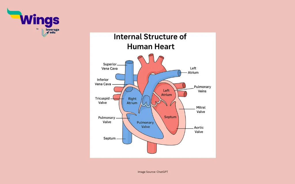
1. Right Atrium
The right atrium is the first chamber that receives deoxygenated blood from the body through two large veins, the superior vena cava and inferior vena cava. Its main function is to collect this blood and pass it into the right ventricle.
2. Right Ventricle
From the right atrium, the deoxygenated blood flows into the right ventricle. The primary function of the chamber is to pump blood to the lungs through the pulmonary artery for oxygenation. The right ventricle’s main function is to send blood to the lungs to get oxygen.
3. Left Atrium
After the blood is oxygenated in the lungs, it returns to the heart via the pulmonary veins and enters the left atrium. This chamber receives the oxygen-rich blood and then passes it to the left ventricle.
4. Left Ventricle
The left ventricle is the thickest and most muscular chamber of the heart. It pumps oxygenated blood to the entire body through the aorta, the body’s largest artery. This ensures all body tissues receive oxygen and nutrients.
Septum
The septum is the thick muscular wall that divides the right and left sides of the heart. It prevents the mixing of oxygenated and deoxygenated blood, ensuring the two circulations remain separate.
Valves of the Heart
The human heart contains four important valves that ensure blood flows in one direction and prevent backflow. These valves play a crucial role in maintaining the efficiency of the pumping action of the heart.
| Heart Valve | Location | Function |
| Tricuspid Valve | Between the right atrium and the right ventricle | Prevents backflow into the right atrium |
| Pulmonary Valve | Between the right ventricle and the pulmonary artery | Controls blood flow from the heart to the lungs |
| Mitral (Bicuspid) Valve | Between the left atrium and the left ventricle | Prevents backflow into the left atrium |
| Aortic Valve | Between the left ventricle and the aorta | Controls blood flow into the aorta and the body |
Coronary Circulation
The heart has its blood supply, known as coronary circulation, which supplies oxygen and nutrients to the heart muscle. This is essential for the continuous working of the heart.
| Blood Vessel, Connected To | Type of Blood Carried | Function |
| Aorta(Left Ventricle) | Oxygenated | Carries oxygen-rich blood from the heart to the entire body |
| Pulmonary Arteries(Right Ventricle) | Deoxygenated | Carry oxygen-poor blood from the heart to the lungs for oxygenation |
| Pulmonary Veins(Left Atrium | Oxygenated | Bring oxygen-rich blood from the lungs back to the heart |
| Vena Cava-Superior & Inferior (Right Atrium) | Deoxygenated | Bring oxygen-poor blood from the body back to the heart |
Check out other Biology questions from here:

 60,000+ students trusted us with their dreams. Take the first step today!